NIH Grant Supports Tiny Experiments Exploring Big Questions
Viewing proteins that are 20 billionths of a meter in size requires a level of precision provided only by a specialized microscope coupled with a spectrograph.
 A new, $385,000 grant from the National Institutes of Health (NIH) will fund an optical microscope that will enable Michael C. Puljung, assistant professor of neuroscience and chemistry, to delve further into questions about the human body at the cellular level.
Puljung pursues the type of science investigations that require meticulous attention to detail. “Using these techniques, I am able to study ion channels ‘at home’ in their native, cellular environment, which is crucial for developing a deeper understanding of their biological role,” said Puljung.
Ion channels are protein pores that generate electrical currents to control heartbeat, muscle contraction, and hormone secretion. Electrical currents result from the movement of millions of charged ions—like sodium and potassium—per second through an open channel. Their dynamic structural changes drive their function.
“To understand these dynamics, I have developed methods to provide high-resolution structural and mechanistic information for intact, functioning channels,” said Puljung. “This will yield specific insights into the mechanism by which severe, disease-causing mutations affect protein function.”
Their dysregulation causes a host of neurological diseases, including epilepsy, migraine, and inherited pain conditions.
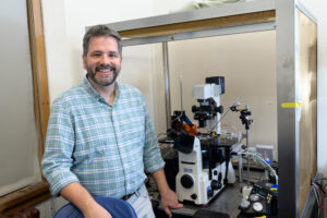
A lab extending off Puljung’s office holds an older version of the precision microscope that will soon be joined by its new companion and double his lab’s capability to explore questions surrounding the functions of proteins in the cell membranes.
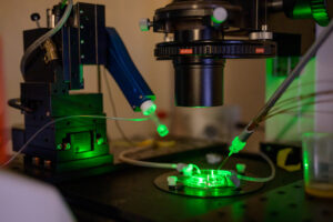
Lifting a dollhouse-sized object attached to the microscope on site, Puljung provides a barely noticeable intake a breath that, unseen to the human eye, attaches a miniscule pipette to a cell’s outer wall and peels off a piece of the membrane. Doing so opens a window to the activity inside the cell.
 Calibrating the tiniest electrical charge to excite the proteins in the cell membrane and shining fluorescent light on the activity generates images that, when interpreted by Puljung or another expert, relate a story.
Prior to investigations, a handheld electrical meter identifies any objects in the lab that may contribute to electrical “noise” that interferes with the readings. The microscope is also located on a table that retains its balance from air pressure to eliminate disturbance from building vibrations.
Creating that setup allows Puljung to marry a cell’s metabolic state to its electrical excitability. “These combined approaches will yield a new appreciation for the role of [specific ion channels] and dysfunction in certain forms of diabetes,” he said.
